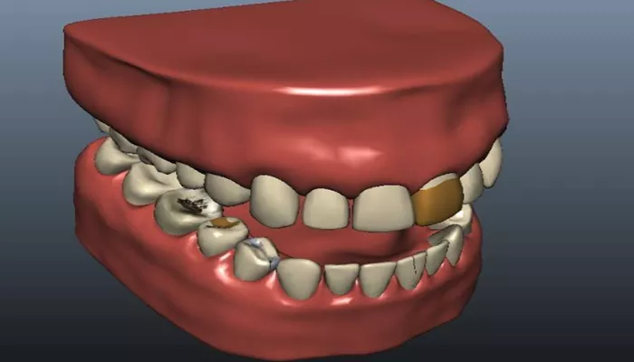
Tooth decay, also known as dental caries, is a common oral health problem. It is a process where the hard tissues of the tooth, mainly the enamel and dentin, are gradually destroyed by acids produced by bacteria in the mouth. X – rays play a crucial role in detecting tooth decay, especially in its early stages when it may not be visible to the naked eye.
Understanding how tooth decay appears on an X – ray is essential for dentists to accurately diagnose and plan appropriate treatment.
Appearance of Initial or Incipient Decay on X – ray
Enamel Decay
In the early stages of tooth decay, when it is mainly affecting the enamel, the decay may appear as a very faint radiolucent area on the X – ray. The enamel is the outermost layer of the tooth and is the hardest tissue in the body. When decay begins, the minerals in the enamel start to be dissolved by the acids. This initial change can be difficult to detect as it is a very subtle alteration in the density of the enamel. The radiolucent area may look like a small, slightly darker patch compared to the surrounding healthy enamel. It might be just a few millimeters in size and could be located near the biting surface of the tooth or in the grooves between the cusps.
For example, in a molar tooth, if decay is starting on the occlusal surface (the part that comes into contact with the opposing tooth during biting), the X – ray may show a tiny, barely noticeable area of decreased density. This is because the acids produced by bacteria are starting to break down the hydroxyapatite crystals in the enamel, causing a change in the way X – rays pass through the tissue.
Dentin Decay
As the decay progresses from the enamel into the dentin, it becomes more visible on the X – ray. Dentin is the layer beneath the enamel and is less dense than the enamel. When decay reaches the dentin, the radiolucent area on the X – ray becomes more pronounced. It may appear as a larger, darker area compared to the initial enamel decay. The shape of the decay in the dentin can vary depending on how the decay has spread. It could be a rounded or oval – shaped area, and it often spreads laterally (sideways) within the dentin.
For instance, if decay has penetrated the enamel and is now in the dentin near the pulp chamber (the central part of the tooth that contains the nerve and blood vessels), the X – ray will show a more distinct area of radiolucency that is closer to the pulp. This is a sign that the decay is getting closer to the vital part of the tooth and may require more urgent treatment.
Advanced Decay on X – ray
Proximity to the Pulp
In cases of advanced decay, the radiolucent area on the X – ray will be very close to or may even be encroaching on the pulp chamber. The pulp is extremely sensitive and important for the health of the tooth. When decay is this close to the pulp, it can cause inflammation and pain. On the X – ray, the decay may look like a large, dark area that is almost touching the outline of the pulp. In some cases, the decay can cause the dentin between the decay and the pulp to become thinned, and this can be seen as a narrow band of less dense tissue on the X – ray.
For example, if a patient has a long – standing cavity that has not been treated, the decay may have spread deep into the tooth, and the X – ray will clearly show the threat to the pulp. This is a critical situation as it may lead to pulpitis (inflammation of the pulp) or even an abscess if left untreated.
Root Decay
Tooth decay can also affect the roots of the tooth, especially in cases where there is gum recession. On the X – ray, root decay appears as a radiolucent area on the root surface. The root is covered by cementum, and when decay affects the root, it can be more difficult to treat. Root decay may look like a small or large area of decreased density along the length of the root. It can be more challenging to detect in its early stages because the root is not as easily visible on X – rays as the crown of the tooth.
For instance, in an older patient with periodontal disease and some gum recession, root decay may be present on the exposed root surfaces. The X – ray may show these areas of decay as faint to more distinct radiolucent patches on the roots.
Conclusion
In conclusion, the appearance of tooth decay on an X – ray varies depending on the stage of decay, the location within the tooth, and the type of decay pattern. Understanding these different appearances is vital for dentists to provide accurate diagnoses and appropriate treatment plans to maintain patients’ oral health.
Related topics:

